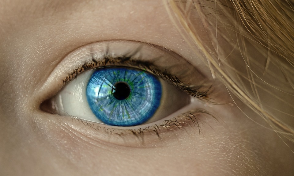
There was a time when the physiology of the eye and its pathologies were mysteries to the humankind. Thankfully, that was a long time ago, and between then and now, scientists and medical experts have come up with thousands of ways to examine the pathophysiology of the eyes. Myopia, hypermetropia, and astigmatism are no longer foreign terms even to the average young adult. In the US, more than 34 million citizens have near-sightedness. In 2015, over 596,000 people with near-sightedness underwent LASIK surgery to improve their eyesight. Due to the advancement of technology, the determination of the most common affliction of the eyes and their treatment has become quick and efficient.
How is the advancement of medical technology help in the diagnosis of complex disorders and diseases of the eye?
How about conditions that are a little more complex than far-sightedness or near-sightedness? What happens when a person suffers from early stages of glaucoma or age-related macular degeneration? Do these patients have to wait till their conditions deteriorate and the symptoms become apparent for concrete diagnosis? Or, are there ways to diagnose pathologies like these without conducting invasive studies?
Advanced medical diagnostic technology brings us super-fast imaging techniques like Optical coherence tomography (OCT) for the diagnosis of ailments like glaucoma. With the help of imaging techniques like Swept-source (SS) OCT, it is now possible to detect the condition at very early stages. It is especially useful in the evaluation of patient’s, who have a family history of the disease. Apart from glaucoma, it can diagnose and determine the progression of macular telangiectasia type 2, retinal diseases like macular degeneration, diabetic retinopathy, macular edema, and retinitis. According to the most recent evaluations of the procedure and its comparisons against the other OCT techniques, the swept source OCT technique offers better clarity in the visualization of the retina, choroid, sclera and vitreous humor. The SS-OCT has improved tissue speeds, deeper tissue penetration, and decreased signal attenuation. It is ideal for studying the structures like the choroid and sclera.
What are the advantages of SS-OCT?
The new swept source OCT technique involves the use of the automated segmentation software. It allows the identification of the retinal layers individually. That makes it possible for the expert to examine the ganglion cell layer (GCL) and the circumcapillary retinal nerve fiber layer (RNFL), from the same scan. The automatic segmentation software can also use the scan to identify different layers including the internal limiting membrane (ILM), inner segment-outer segment junction (IS/OS), inner plexiform layer (IPL), RPE, Bruch’s membrane and choroid. The ability of the technique in coordination with the software to generate multiple thickness maps of the different cross sections is primarily instrumental in modern diagnostics.
- One of the most significant advantages of the SS-OCT is its high-speed imaging technique. The high imaging speed allows high-resolution imaging irrespective of the patient’s eye movements. This technique can negate the effects of the eye movements while generating the high-res images. In contrast to the SD-OCT, the SS-OCT technique uses an invisible light source, which proves to be less distracting to the patient during the time of the test.
- Another advantage is the use of long-wavelength invisible light enables the development of clear images of the lamina cribrosa and the choroid. This type of wavelength scatters less in the retinal pigment epithelium (RPE). Thus the long-wavelength enables high-quality imaging of the inner structures that lay deep within the eye.
Both of these advantages contribute to the ease of confirmed diagnosis of glaucoma in the subjects.
How does SS-OCT help in the diagnosis and treatment of glaucoma?
Improving the understanding of the disease mechanisms, detection of the diseases and the identification of the novel risk factors. SS-OCT is bringing a new wave of diagnostic ease and accuracy in open-angle glaucoma. The ability of the imaging technique to form multiple layered images from a single scan, allows the expert to see the RNFL defects towards the macula, the GCL+ thickness and a combination of the prior ones to generate a GCC map. The SS-OCT technique creates a complete summary of the prognosis of the disease.
A recent study in the University of San Diego, the experts, studied 172 eyes with glaucoma, and 96 healthy eyes acted as the control. It was the Diagnostic Innovations and Glaucoma Study (DIGS) that defined glaucoma as the presence of two or more abnormal visual fields on the stereophotographs in the masked gradings. The early results of this study were promising and in coordination with the results from perimetry tests that the experts conducted on the subjects consecutively. SS-OCT or Deep Range Imaging OCT is a reliable tool in the detection of glaucoma.
Another significant advantage of leveraging the SS-OCT tech in the diagnosis of glaucoma is the close examination of the retinal ganglion cell (RGC) axons. It is the preliminary site of damage in glaucoma cases. It is a 3-dimensional meshwork of fenestrated capillaries, sheets of the scleral collagenous tissue and variegated elastic fibers. In a glaucomatous eye, the lamina cribrosa can undergo significant morphological changes that early histological, in-vitro studies have shown. Now it is possible to observe these changes and stage the disease from data in real-time without any invasive procedures. SS-OCT gives the world a way to diagnose open-angle glaucoma in the population before the disease starts showing significant symptoms that can result in a blurry vision to a sudden loss of eyesight.
What’s the verdict on SS-OCT?
This third-generation OCT technology is a potent diagnostic tool. It is quick and accurate. While convention OCT is much better than the technologies that came before it, the modern SS-OCT is much faster, and detailed than any existing OCT technique. The third-generation SS-OCT is undoubtedly versatile, and it can help in the qualitative and quantitative assessment of the retinal nerve damage, choroidal damage and studies of the lamina cribrosa. It can further our understanding of the different mechanisms of the eye, the axonal damage and the development of additional risk factors that may arise from it.
Recent Posts
- Castor Oil For Better Hair Growth: Is It Myth Or Fact?
- Exploring the Differences Between Sermorelin, Ipamorelin, Ibutamoren, GHRP2, and GHRP6: Understanding Their Role in Human Growth Hormone Regulation
- Unraveling the Mystery: Understanding the Causes and Prognosis of Ventricular Tachycardia Without Apparent Heart Disease
- Understanding Grandparents’ Rights in Oklahoma: Navigating Visitation and Legal Protections
- 10 Reasons to Consider Hypnotherapy for Your Health

